- Home
- Up to XII Students
- Decide Stream For XI & XII
- Career Options
- Entrance Preparation
- Sample Papers
- Entrance Exams after 12th
- International Entrance Exams To Study Abroad
- Foreign University Comparison
- Apply to Foreign Universities
- General Preparation For Students Going To Study Abroad
- Write A Report
- Write an Article
- Write An Essay
- Important Dates
- UG & PG
- Tech Tips
- Mock Test
- GROOMING
- HOBBIES
- K PLUS
- Working Professional
- OTHERS
DTI AND fMRI: THE IMAGING MODALITY FOR BRAIN TUMORS
Divya,
Assistant Professor, Department of Computer Science
divya.awasthi@kalingauniversity.ac.in, jangid.divya@gmail.com
Imaging plays a crucial role in the assessment of patients with brain tumours. A number of imaging modalities such as Computed Tomography Scan (CT-Scan), Magnetic Resonance Imaging (MRI), Ultrasound, Positron Emission Tomography scan (PET) and Diffusion tensor imaging (DTI) .The two most significant and widely used imaging modalities are CT scan and MRI Scan. They significantly influence patient care with their analysis parameters. The identification and assessment of brain neoplasms has substantially improved with the technological advancement of CT and MRI, the usefulness of contrast material in the imaging of brain tumours, and the advent of new imaging techniques.

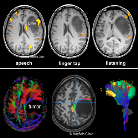
Figure 1 MRI and DTI
Brain
activity may be visualised in great detail using MRI and functional MRI. It is
employed to pinpoint precisely which region of the brain controls important
processes including thought, speech, vision, movement, and sensation.
Additionally, it can display how Alzheimer’s disease, trauma, or a stroke
affects brain function. Using a technique called Diffusion Tensor Imaging
(DTI), it is possible to track the path that water takes through the brain’s
white matter pathways. The brain’s white-matter pathways, which connect various
regions, act like a safeguarded during surgery. The white matter fibres that
link various areas of the brain are found using DTI. Despite the fact that
traumatic brain injury has been the focus of most DTI research, it also has
other uses, such as helping to diagnose, predict outcomes, categorise stroke,
brain tumours, neurodegenerative diseases, developmental disorders,
neuropsychiatric disorders, movement disorders, and neurogenetic developmental
disorders. Numerous measurements that are computed using DTI can offer
quantitative power. Fractional anisotropy (FA) is one of the DTI parameters
that are most frequently utilized for Brain tumour diagnosis; others are Radial
Diffusivity (perpendicular), Axial Diffusivity (parallel), Average Diffusivity
and Apparent Diffusion Coefficient (ADC). FA is highly sensitive to changes in
microstructure and as a summary quantifies the directionality of diffusivity,
but it is not always able to pinpoint the exact origin of a change. The region
of interest (ROI) approach, whole-brain analysis (Voxel-Based analysis), and
tract-based spatial statistics can all be used to compute FA values. Due to its
automation and capacity to examine more tracts, whole-brain analysis is
becoming more and more popular. The ROI approach, in which the regions to be
examined are marked by a technician before being examined by a computer, is
still dependable and repeatable.
References:
·
Drevelegas, A. (2002). Imaging Modalities in Brain Tumors.
In: Drevelegas, A. (eds) Imaging of Brain Tumors with Histological
Correlations. Springer, Berlin, Heidelberg.
https://doi.org/10.1007/978-3-662-04951-8_2
·
Ranzenberger LR, Snyder T. Diffusion Tensor Imaging. [Updated 2022 Jul
26]. In: StatPearls [Internet]. Treasure Island (FL): StatPearls Publishing; 2023
Jan-.
·
Yahya M A Mohammed and others, A survey of methods for brain tumor
segmentation-based MRI images, Journal of Computational Design
and Engineering, Volume 10, Issue 1, February 2023, Pages
266–293, https://doi.org/10.1093/jcde/qwac141.
·
Işın, Ali &
Direkoglu, Cem & Sah, Melike. (2016). Review of MRI-based Brain Tumor Image
Segmentation Using Deep Learning Methods. Procedia Computer Science. 102.
317-324. 10.1016/j.procs.2016.09.407.

Kalinga Plus is an initiative by Kalinga University, Raipur. The main objective of this to disseminate knowledge and guide students & working professionals.
This platform will guide pre – post university level students.
Pre University Level – IX –XII grade students when they decide streams and choose their career
Post University level – when A student joins corporate & needs to handle the workplace challenges effectively.
We are hopeful that you will find lot of knowledgeable & interesting information here.
Happy surfing!!
- →
-
Free Counseling!
